
 Researchers at the ARC Centre for Excellence in Nanoscale BioPhotonics (CNBP) discovered an exciting new method that could make it possible to use 3D microscopy to view hard-to-reach areas of the human body. This method uses fiber optic bundles to miniaturize a type of 3D imaging called “light field imaging.” Taken to extreme new levels, this imaging could make in-body use possible.
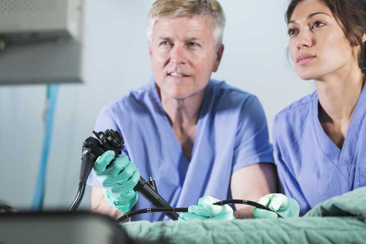
This method could find widespread use in diagnostic procedures called optical biopsies. In these biopsies, suspicious body tissue is investigated using medical endoscopic procedures.
Until now, light field imaging could be performed only with bulky hardware such as camera arrays or modified consumer cameras. Instead of trying to shrink existing equipment, the researchers realized that fiber optic bundles already used routinely in microendoscopy were actually suitable light field imaging devices.
Fiber optic bundles are groups of tens of thousands of microscopic optical fibers. Each fiber in the bundle acts like a pixel in a camera. The result produced is a 2D image that is transmitted through the fiber bundle.
Along with recording a 2D picture, light field imaging systems also measure the incoming angles of all light rays in the picture. With this information, doctors can map the picture in stereo 3D in the same way that humans perceive depth. According to the researchers, the primary challenge will be how to record this angular light ray dimension that is often hard to capture.
According to Dr. Antony Orth, project lead and Research Fellow at the RMIT University node of the CNBP, “The key observation we made is that light ray orientation information is actually transmitted by the fiber optic bundles to the microendoscope. You just need to know what to look for and how to decode it.”
Dr. Orth believes that, given the right mathematical framework, researchers can decode the patterns, transform them into a light field, and do incredible things such as refocusing, depth mapping and visualizing the image in stereo 3D. He believes that this light field technology could potentially bring an entirely new depth dimension into optical biopsies. This capability would let doctors examine suspect tissue without removing a sample from the patient.
The research group is meeting with physicians to discuss how to test this technique in medical clinics and to also identify the medical procedures most likely to benefit from 3D visualization in the microscale.
Tags: endoscope, fiber optics, medical imaging